New technology unwraps mummies' ancient mysteries
LONDON -- Our fascination with mummies never gets old. Now the British Museum is using the latest technology to unwrap their ancient mysteries. Scientists at the museum have used CT scans and sophisticated imaging software to go beneath the bandages, revealing skin, bones, preserved internal organs -- and in one case a brain-scooping rod left inside a skull by embalmers...
LONDON -- Our fascination with mummies never gets old. Now the British Museum is using the latest technology to unwrap their ancient mysteries.
Scientists at the museum have used CT scans and sophisticated imaging software to go beneath the bandages, revealing skin, bones, preserved internal organs -- and in one case a brain-scooping rod left inside a skull by embalmers.
The findings go on display next month in an exhibition that sets eight of the museum's mummies alongside detailed three-dimensional images of their insides and 3-D printed replicas of some of the items buried with them.
Bio-archaeologist Daniel Antoine said Wednesday that the goal is to present these long-dead individuals "not as mummies but as human beings."
Mummies have been one of the British Museum's biggest draws ever since it opened in 1759. Director Neil MacGregor said 6.8 million people visited the London institution last year, "and every one asked one of my colleagues, 'Where are the mummies?'"
The museum has been X-raying its mummies since the 1960s, but modern CT scanners give a vastly sharper image. Just like live patients, the mummies chosen for the exhibition were scanned at London hospitals -- though they were wheeled in after hours.
Volume graphics software, originally designed for car engineering, was then used to put flesh on the bones of the scans -- showing skeletons, adding soft tissue, exploring the nooks and cavities inside.
The eight mummies belong to individuals who lived in Egypt or Sudan between 3,500 B.C. and 700 A.D. They range from poor people naturally preserved in sand -- the cheapest burial option -- to high-ranking Egyptians given elaborate ceremonial funerals.
"You got what you paid for, basically," said museum mummy expert John Taylor. "There were different grades of mummification."
Embalmers were exceptionally skilled, extracting the brain of the deceased through the nose, although they sometimes made mistakes.
The museum's scientists were thrilled to discover a spatula-like probe still inside one man's skull, along with a blob of brain.
"The tool at the back of the skull was quite a revelation, because embalmers' tools are something that we don't know much about," Taylor said. "To find one actually inside a mummy is an enormous advance."
The man, who died around 600 B.C., also had painful dental abscesses that might have killed him. Another mummy, a woman who lived in Sudan around 700 A.D. was a Christian with a tattoo of the Archangel Michael's name on her inner thigh.
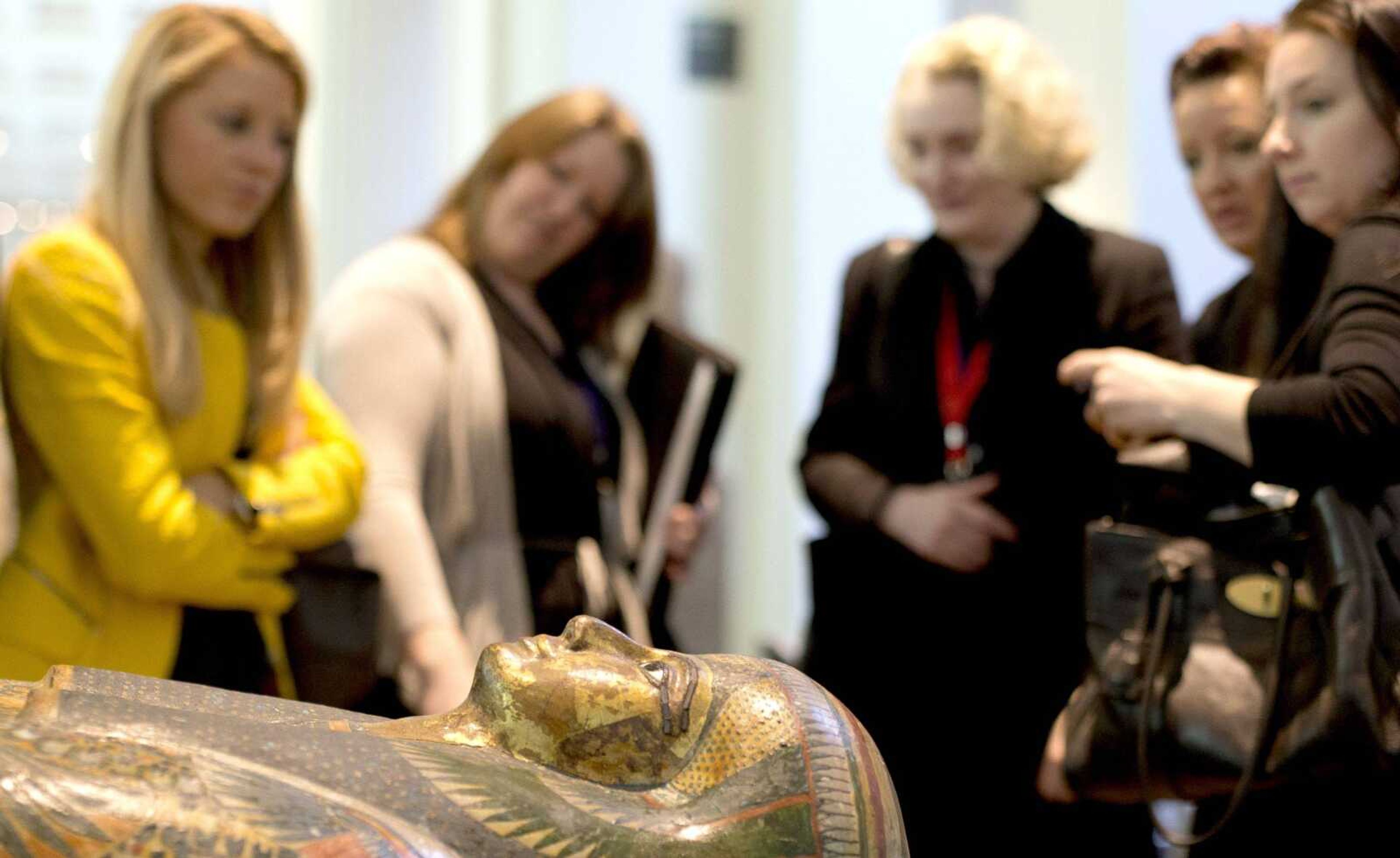
The star of the show is Tamut, a temple singer from a family of high-ranking priests who died in Thebes around 900 B.C. Her brightly decorated casket, covered in images of birds and gods, has never been opened, but the scans have revealed in extraordinary detail her well-preserved body, down to her face and short-cropped hair.
Tamut was in her 30s or 40s when she died, and had calcified plaque inside her arteries -- a sign of a fatty diet and high social status. She may well have died from a heart attack or stroke.
Several amulets carefully are arranged on her body, including a figure of a goddess with its wings spread protectively across her throat. It's even possible to see beeswax figurines of gods placed inside her chest to protect the internal organs in the afterlife.
"The clarity of the images is advancing very rapidly," Taylor said. "As the technology advances, we have hopes that we may be able to read even hieroglyphic inscriptions on objects inside mummies."
MacGregor said the museum plans eventually to scan all 120 of its Egyptian and Sudanese mummies, and to reveal even more about their lives.
"Come back in another five years and you will hear Tamut sing," he said.
Connect with the Southeast Missourian Newsroom:
For corrections to this story or other insights for the editor, click here. To submit a letter to the editor, click here. To learn about the Southeast Missourian’s AI Policy, click here.


